- +1 858 909 0079
- +1 858 909 0057
- [email protected]
- +1 858 909 0079
- [email protected]

The interaction between avidin (or streptavidin) and biotin exhibits one of the highest known non-covalent interactions (Fig.1). Avidin, streptavidin, monomeric avidin, and their analogs have become powerful tools for probes and affinity ligands for various biochemical assays, diagnosis, affinity purification, and drug delivery applications.

Avidin is a biotin-binding glycosylated protein derived from chicken white. It comprises four 128 amino acid subunits, each binding one biotin molecule over a wide range of pH and temperature. Since avidin can be chemically modified with little to no effect on its function and is easily isolated from chicken egg whites, it helps detect biotinylated molecules in various conditions.
Streptavidin is a tetrameric biotin-binding protein that originates from Streptomyces avidin and has a mass of 60 kDa. It has no carbohydrate and lower pI, giving a lower degree of nonspecific binding, making streptavidin an ideal reagent choice for many detection systems.
●
High affinity
●
High stability
The biotin-binding complex is exceptional stability and can resist Extreme pH, temperature (2 °C and 40 °C), harsh organic solvents, denaturing agents such as guanidinium chloride, detergents such as SDS and proteolytic enzymes.
●
Outstanding selectivity and specificity
The biotin and streptavidin or avidin interaction are highly specific, ensuring low nonspecific binding.
●
High sensitivity
High stability and specificity ensure high detection sensitivity.
●
Very flexibility
The small size and remarkable stability of biotin are exceptionally suitable for relatively easy incorporation into various molecules or specific locations in molecules with minimal perturbation to the structure and function of the conjugated biomolecules.
Compared to other non-covalent interactions, the biotin-binding system provides tremendous benefits. It has become a versatile platform for amplifying weak signals, flow cytometry, Western blotting, ELISA, FLISA, IHC, immunofluorescence microscopy, etc. However, currently, the applications of the biotin-binding systems are mainly based on the traditional affinity chromatography matrices such as beaded agarose resin or column. Their procedures are tedious, time-consuming, and unable to handle tiny samples such as cancer-cell targeting and single-cell analysis. We developed an extremely efficient magnetic affinity system to overcome these limitations.
Magnetic beads (particles) are an entirely different type of solid support matrices from beaded agarose or other porous resins. They are much smaller (typically 1-5 µm diameter), thus providing larger surface areas for a high density of ligand immobilization. The beads are manufactured using nanometer-scale superparamagnetic iron oxide as core and entirely encapsulated by a high purity silica shell, ensuring no leaching problems with the iron oxide. The pure inert silica makes less nonspecific binding.
1.
BcMag™ Streptavidin Magnetic Beads
The Magnetic particle is a highly uniform and superparamagnetic microsphere coated with a high density of high purity (>97%) streptavidin on the surface. The beads are manufactured using nanometer-scale superparamagnetic iron oxide as core and entirely encapsulated by a high purity silica shell, ensuring no leaching problems with the iron oxide. The microspheres are specifically designed and tested for applications in immunoprecipitation, cell sorting, and rapid single-step capture of the target molecules such as DNA, RNA, antibody, or protein from cell lysates or hybridization reactions.
Learn More
2.
BcMag™ Cleavable Streptavidin Magnetic Beads
BcMag™ Cleavable Streptavidin Magnetic Beads are uniform superparamagnetic microspheres coated with high purity of (>97%) streptavidin. Because streptavidin is linked to the beads (particles) via a cleavable disulfide linker, reducing agents such as DTT or -mercaptoethanol can cleave and separate the biotinylated molecule-streptavidin complex from the beads rather than using harsh elution reagents such as 8M guanidine or SDS detergent after affinity purification. The microspheres have been specifically designed and evaluated for use in immunoprecipitation, cell sorting, and quick single-step capture of biotinylated molecules from cells such as DNA, RNA, antibodies, or proteins.

Learn More
3.
BcMag™ Streptavidin Terbium Fluorescent Magnetic Beads
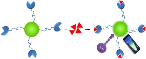
The green fluorescent magnetic resin is highly-fluorescent, uniform, and superparamagnetic microspheres coated with streptavidin. The microspheres combine all the advantages of a unique streptavidin biotin-binding system, time-resolved fluorescent dyes, and magnetic properties to perform highly sensitive assays. The beads are manufactured using nanometer-scale superparamagnetic iron oxide and terbium metal as core and entirely encapsulated by a high purity silica shell, ensuring no leaching problems with the iron oxide and terbium metal.

Learn More
4.
BcMag™ Streptavidin Europium Fluorescent Magnetic Beads
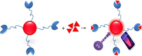
The red fluorescent magnetic beads are highly-fluorescent and uniform superparamagnetic microspheres coated with streptavidin. The microspheres combine all the advantages of a unique streptavidin biotin-binding system, time-resolved fluorescent dyes, and magnetic properties to perform highly sensitive assays. The intensely red fluorescent microspheres (particles) are manufactured using nanometer-scale iron oxide and europium metal as core and entirely encapsulated by a high purity silica shell, typically ensuring no leaching problems with the iron oxide and terbium metal.

Learn More
5.
BcMag™ Monomer Avidin Magnetic Beads

Learn More
7.
BcMag™ Avidin Europium Fluorescent Magnetic Beads
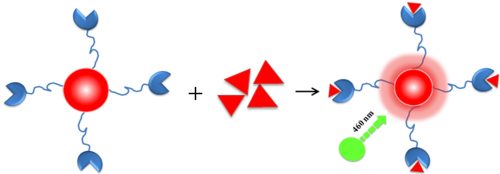
The super bright red fluorescent magnetic beads are highly-fluorescent uniform superparamagnetic microspheres coated with a high density of avidin. The microspheres combine all the advantages of a unique avidin biotin-binding system, time-resolved fluorescent dyes, and magnetic properties to perform highly sensitive assays. The beads are manufactured using nanometer-scale superparamagnetic iron oxide and Terbium as core and entirely encapsulated by a high purity silica shell, ensuring no leaching problems with the iron oxide and terbium metal.
Learn More
8.
BcMag™ Avidin Ruthenium Fluorescent Magnetic Beads
The Beads are highly-fluorescent uniform superparamagnetic microspheres coated with a high density of avidin. The microspheres combine all the advantages of a unique avidin biotin-binding system, time-resolved fluorescent dyes, and magnetic properties to perform highly sensitive assays. The beads are manufactured using nanometer-scale superparamagnetic iron oxide and ruthenium metal as core and entirely encapsulated by a high purity silica shell, ensuring no leaching problems with the iron oxide and terbium metal.

Learn More
9.
BcMag™ Streptavidin Ruthenium Fluorescent Magnetic Beads
BcMag™ Streptavidin Ruthenium Fluorescent Magnetic Beads are highly-fluorescent, uniform, and superparamagnetic microspheres coated with streptavidin protein. The microspheres combine all the advantages of a unique avidin biotin-binding system, time-resolved fluorescent dyes, and magnetic properties to perform highly sensitive assays. The beads are manufactured using nanometer-scale superparamagnetic iron oxide and ruthenium metal as core and entirely encapsulated by a high purity silica shell, ensuring no leaching problems with the iron oxide and terbium metal.
Learn More
10.
His-tagged recombinant streptavidin
The highly pure (>95%) recombinant streptavidin protein with 6x His-tag at N-terminus was produced in E.coli and purified by Ni resin. It is a single, non-glycosylated polypeptide chain and has a molecular mass of 18 kDa. The recombinant streptavidin is widely used to detect biotinylated biomolecules or as probes in various applications such as Western blotting, ELISA, immunohistochemistry, and fluorescence imaging.
1.
Dong H, An X, Xiang Y, Guan F, Zhang Q, Yang Q, Sun X, Guo Y. Novel Time-Resolved Fluorescence Immunochromatography Paper-Based Sensor with Signal Amplification Strategy for Detection of Deoxynivalenol. Sensors (Basel). 2020 Nov 18;20(22):6577. doi: 10.3390/s20226577. PMID: 33217912; PMCID: PMC7698798.
2.
Xiao X, Haushalter JP, Kotz KT, Faris GW. Cell assay using a two-photon-excited europium chelate. Biomed Opt Express. 2011;2(8):2255-2264. doi:10.1364/BOE.2.002255
3.
Hemmilä I, Dakubu S, Mukkala VM, Siitari H, Lövgren T. Europium as a label in time-resolved immunofluorometric assays. Anal Biochem. 1984 Mar;137(2):335-43. doi: 10.1016/0003-2697(84)90095-2. PMID: 6375455
4.
Cho U, Chen JK. Lanthanide-Based Optical Probes of Biological Systems. Cell Chem Biol. 2020 Aug 20;27(8):921-936. doi: 10.1016/j.chembiol.2020.07.009. Epub 2020 Jul 30. PMID: 32735780; PMCID: PMC7484113.
5.
Mizukami S, Tonai K, Kaneko M, Kikuchi K. Lanthanide-based protease activity sensors for time-resolved fluorescence measurements. J Am Chem Soc. 2008 Nov 5;130(44):14376-7. doi: 10.1021/ja800322b. Epub 2008 Oct 8. PMID: 18839953.
6.
Johansson MK, Cook RM, Xu J, Raymond KN. Time gating improves sensitivity in energy transfer assays with terbium chelate/dark quencher oligonucleotide probes. J Am Chem Soc. 2004 Dec 22;126(50):16451-5. doi: 10.1021/ja0452368. PMID: 15600347.
7.
Chivers CE, Koner AL, Lowe ED, Howarth M. How the biotin-streptavidin interaction was made even stronger: investigation via crystallography and a chimaeric tetramer. Biochem J. 2011;435(1):55-63. doi:10.1042/BJ20101593
8.
Dundas CM, Demonte D, Park S. Streptavidin-biotin technology: improvements and innovations in chemical and biological applications. Appl Microbiol Biotechnol. 2013 Nov;97(21):9343-53. doi: 10.1007/s00253-013-5232-z. Epub 2013 Sep 22. PMID: 24057405.
9.
Lakshmipriya T, Gopinath SC, Tang TH. Biotin-Streptavidin Competition Mediates Sensitive Detection of Biomolecules in Enzyme Linked Immunosorbent Assay. PLoS One. 2016 Mar 8;11(3):e0151153. doi: 10.1371/journal.pone.0151153. PMID: 26954237; PMCID: PMC4783082.
10.
Yang H, Zhang Q, Liu X, Yang Y, Yang Y, Liu M, Li P, Zhou Y. Antibody-biotin-streptavidin-horseradish peroxidase (HRP) sensor for rapid and ultra-sensitive detection of fumonisins. Food Chem. 2020 Jun 30;316:126356. doi: 10.1016/j.foodchem.2020.126356. Epub 2020 Feb 4. PMID: 32045810.
Magnetic Beads Make Things Simple