- +1 858 909 0079
- +1 858 909 0057
- support@bioclone.net
- +1 858 909 0079
- support@bioclone.net

Products
Cat. No.
Product Name
Unit Size
Order
Specification
Beads Size
~ 2.5 μm diameter
Number of Beads
~ 1.47 x 10⁸ beads/mg
Magnetization
~40-45 EMU/g
Type of Magnetization
Superparamagnetic
Concentration
Binding Capacity
Biotinylated BSA / ml of Beads
>1mg/ml
Biotinylate single-stranded oligonucleotides
~ 2,000 pmoles /ml
Storage
Ship at room temperature. Store at 4°C. Do not freeze.
Shipping conditions: At ambient temperature
Handling and Storage: Store the kit components according to the table above on arrival.
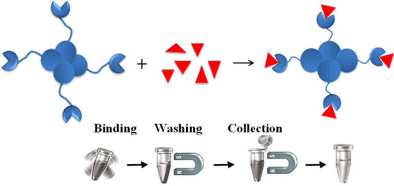
BcMag™ Streptavidin Magnetic Bead is a highly uniform and superparamagnetic microsphere coated with a high density of high purity (>97%) streptavidin on the surface. The beads are manufactured using nanometer-scale superparamagnetic iron oxide as core and entirely encapsulated by a high purity silica shell, ensuring no leaching problems with the iron oxide. The microspheres are specifically designed and tested for applications in immunoprecipitation, cell sorting, and rapid single-step capture of the target molecules such as DNA, RNA, antibody, or protein from cell lysates or hybridization reactions.
The interaction between streptavidin (or avidin n) and biotin exhibits one of the highest known non-covalent interactions). Avidin, streptavidin, monomeric avidin, and their analogs have become powerful tools for probes and affinity ligands for a wide range of applications in biochemical assays, diagnosis, affinity purification, and drug delivery. Streptavidin is a tetrameric biotin-binding protein that originates from Streptomyces avidinii and has a mass of 60,000 daltons. Streptavidin has no carbohydrate and lower pI, giving a lower degree of nonspecific binding, making streptavidin an ideal reagent choice for many detection systems.
Advantages and benefits of using streptavidin biotin-binding systems
●
High affinity
●
High stability
The biotin-binding complex is exceptional stability and can resist Extreme pH, temperature (2 °C and 40 °C), harsh organic solvents, denaturing agents such as guanidinium chloride, detergents such as SDS and proteolytic enzymes.
●
Outstanding selectivity and specificity
The interaction of the biotin and streptavidin is highly specific, ensuring low nonspecific binding.
●
High sensitivity
High stability and specificity ensure high detection sensitivity.
●
Very flexibility
The small size and remarkable stability of biotin are proven to be exceptionally suitable for relatively easy incorporation into a variety of molecules such as synthetic polymers, fluorophores, small molecules, or into specific locations in biomolecules, that imposes minimal perturbation to the structure and function of the conjugated biomolecules.
Advantages and benefits of using streptavidin magnetic microspheres
●
Quick, Easy, and one-step high-throughput procedure: eliminates Columns or filters, or a laborious repeat of pipetting or centrifugation
●
High binding capacity
●
Exhibits low nonspecific binding
●
Scalable – Easily adjusts for sample size and automation
Materials Required
●
Binding Buffer: 1x PBS, 0.1% BSA, pH 7.4
●
Magnetic Rack (for manual operation)
Based on sample volume, the user can choose one of the following Magnetic Racks:
– BcMag™ Magnetic Rack-6 for holding six individual 1.5 ml centrifuge tubes (Cat. No. MS-02);
– BcMag™ Magnetic Rack-24 for holding twenty-four individual 1.5-2.0 ml centrifuge tubes (Cat. No. MS-03);
– BcMag™ Magnetic Rack-50 for holding one 50 ml centrifuge tube, one 15 ml centrifuge tube, and four individual 1.5 ml centrifuge tubes (Cat. No. MS-04);
– BcMag™ Magnetic Rack-96 for holding a 96 ELISA plate or PCR plate (Cat. No. MS-05).
Procedure
Note:
●
This protocol is a general affinity purification procedure. To obtain the best results, each user should determine the optimal working conditions for the purification of the individual target protein.
●
We strongly recommended titration to optimize the number of beads used for each application based on the amount of the target biotinylated molecules in the crude sample based on the beads binding capacity. Too many magnetic beads used will cause higher backgrounds, while too few beads used will cause lower yields.
1.
Transfer the optimal amount of the beads to a centrifuge tube. Place the tube on the magnetic rack for 1-3 minutes. Remove the supernatant while the tube remains on the rack.
2.
Remove the tube and wash the beads with 5-bed volumes of PBS buffer by vortex for 30 seconds. Leave the tube at room temperature for 1-3 minutes. Place the tube on the magnetic rack for 1-3 minutes. Remove the supernatant while the tube remains on the rack.
3.
Repeat step 2 two times.
4.
Add washed beads to the crude sample containing target molecules and incubate at room temperature or desired temperature for 1-2 hours (Lower temperature require longer incubation time).
Note: Strongly recommended to perform a titration to optimize incubation time. More prolonged incubation may cause higher background.
5.
Note: Adding a higher concentration of salts, nonionic detergent, and reducing agents may reduce the nonspecific background. For example, the addition of NaCl (up to 1-1.5 M), 0.1-0.5% nonionic detergents such as Triton X 100 or Tween 20 to the washing buffer.
Questions and Answers
1.
How is it possible to break the bond between biotin and streptavidin?
The chemical bond between biotin and streptavidin is incredibly strong. It can break under denaturing conditions. Many applications do not require the separation of biotin from streptavidin. However, if the user needs to separate the biotin from streptavidin, follow the following suggestions:
●
Protein or antibody
To elute the bound biotinylated protein or antibody from the beads, add the appropriate amount of Elution Buffer (0.1 M Glycine-HCl, pH 2.5), incubate for 0.5-5 min at room temperature, collect the magnetic beads by a magnetic rack and transfer the supernatant to a fresh tube. Immediately neutralize the supernatant to pH 7.0 by adding 1/10 volume of Neutralization Buffer (1.0 M Tris-HCl, pH 9.0).
●
DNA
Long nucleic acids used as biotinylated probes are not recommended due to difficulty in elution. For short biotinylated oligos, the user can try the following method:
a. To elute the bound biotinylated oligo from the beads, add an appropriate amount of Elution Buffer (10 mM EDTA, pH 8.2, 95% formamide), incubate at 65º C for 2 min.
b. Collect the magnetic beads by the magnetic rack and transfer the supernatant to a fresh tube.
2.
How can I elute the bound nonbiotinylated sample from the Magnetic Beads?
●
Protein or antibody
If a non-biotinylated protein or antibody interacts with a biotinylated antibody or protein, add an appropriate amount of Elution Buffer (0.1 M Glycine-HCl, pH 2.5), incubate for 30 sec-5 min at room temperature, collect the magnetic beads by the magnetic rack and transfer the supernatant to a fresh tube. Immediately neutralize the supernatant to pH 7.0 by adding 1/10 volumes of Neutralization Buffer (1.0 M Tris-HCl, pH 9.0).
●
DNA or RNA
1.
Chivers CE, Koner AL, Lowe ED, Howarth M. How the biotin-streptavidin interaction was made even stronger: investigation via crystallography and a chimaeric tetramer. Biochem J. 2011;435(1):55-63. doi:10.1042/BJ20101593
2.
Dundas CM, Demonte D, Park S. Streptavidin-biotin technology: improvements and innovations in chemical and biological applications. Appl Microbiol Biotechnol. 2013 Nov;97(21):9343-53. doi: 10.1007/s00253-013-5232-z. Epub 2013 Sep 22. PMID: 24057405.
3.
Lakshmipriya T, Gopinath SC, Tang TH. Biotin-Streptavidin Competition Mediates Sensitive Detection of Biomolecules in Enzyme Linked Immunosorbent Assay. PLoS One. 2016 Mar 8;11(3):e0151153. doi: 10.1371/journal.pone.0151153. PMID: 26954237; PMCID: PMC4783082.
4.
Yang H, Zhang Q, Liu X, Yang Y, Yang Y, Liu M, Li P, Zhou Y. Antibody-biotin-streptavidin-horseradish peroxidase (HRP) sensor for rapid and ultra-sensitive detection of fumonisins. Food Chem. 2020 Jun 30;316:126356. doi: 10.1016/j.foodchem.2020.126356. Epub 2020 Feb 4. PMID: 32045810.
Magnetic Beads Make Things Simple