- +1 858 909 0079
- +1 858 909 0057
- support@bioclone.net
- +1 858 909 0079
- support@bioclone.net

Products
Cat. No.
Product Name
Unit Size
Order
Fluorophore
Excitation
(nm)
Emission
(nm)
Fluorescence lifetime
(Ԏ) (µsec)
Stokes shifts
(nm)
Selection of
Emission Filter
Green
320
545
1050
220
545/40
Fluorescence Dye Properties
Fluorophore
Terbium (Tb3+)
Green
320
545
Fluorescence lifetime (Ԏ) (µsec)
1050
Stokes shifts
(nm)
220
Selection of
Emission Filter
545/40
Specification
Bead Size
2.5μm diameter; 5μm diameter
Number of Beads
~ 10 x 107 beads/mg (2.5μm)
~ 5 x 10⁷ beads /mg (5μm)
Magnetization
~40-45 EMU/g
Type of Magnetization
Superparamagnetic
Concentration
10 mg/ml (10mM Tris, 0.15 M NaCl, 0.1% BSA,1 mM EDTA, pH 7.4)
Binding Capacity
~ 1 mg IgG/ml of Beads
Storage
Ship at room temperature. Store at 4°C. Do not freeze.
BcMag™ Protein G Terbium Fluorescence Magnetic Beads are Time-Resolved Fluorescence (TRF) magnetic microspheres covalently coupled with Protein G on the surface. The beads are manufactured using nanometer-scale superparamagnetic iron oxide and terbium metal as core and entirely encapsulated by a high-purity silica shell, ensuring no leaching problems with the iron oxide and europium metal. The microspheres combine the benefits of a novel avidin biotin-binding method, time-resolved Fluorescence dyes, and magnetic characteristics to perform very sensitive assays.
Despite being widely utilized over the past few decades, conventional fluorophores still face several limitations that hinder their effectiveness and applicability. These limitations include a narrow excitation band that can lead to higher background signals, a smaller Stokes shift that may result in self-quenching, sensitivity to environmental factors such as pH, temperature, metallic ion concentration, and solvent polarity, low fluorescence intensity that may not be sufficient for detecting a single biomolecule, fluorescence intermittency (blinking) that can affect molecule detection processes, and a tendency to aggregate due to hydrophobicity.
BcMag™ TR-FRET Assay, in contrast to typical FRET (Förster Resonance Energy Transfer) assays, uses time-resolved Fluorescence magnetic beads (BcMag™ TR-Magnetic Beads) as the donor fluorophore. The donor and acceptor can be two proteins, two DNA strands, an antigen, an antibody, or a ligand and its receptor. After a reasonable time delay (usually 50 to 100 s), a signal is generated by fluorescence resonance energy transfer between a donor and an acceptor molecule when they are close and monitored in a time-resolved way. In BcMag™ TR-FRET Assay, a trace amount of analytes can be easily enriched from the complex by TR-Magnetic Beads, resulting in higher sensitivity. This assay practically eliminates all fluorescence backgrounds caused by the sample and plastic microplate, as well as by direct acceptor excitation. As a result, the signal-to-noise ratios of the BcMag™ TR-FRET Assay are very high, and the background is quite low. Furthermore, the assay does not need washing steps. BcMag™ TR-FRET Assay offers substantial advantages to bioassays in high throughput screening, such as assay flexibility, dependability, increased assay sensitivity, higher throughput, and fewer false positive/false negative results.
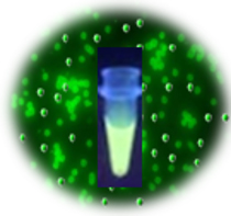
Terbium cryptate fluorophore is an efficient Fluorescence label due to its distinct specific properties. It is excited at 320nm and emits green fluorescence at 545nm, with a long fluorescence lifetime (1050 μsec) and large stokes shifts (220 nm). By taking advantage of these properties, time-resolved fluorescence measurement can dramatically reduce the fluorescence background from the sample and increase the signal-to-noise ratio to offer detectability better than one order of magnitude than conventional Fluorescence dyes. BcMag™ Protein G Terbium Fluorescence Magnetic Beads are excellent donors used in TR-FRET assays.
1.
Mix the antibody-conjugated donor beads with the cell lysates and incubate them with continuous rotation for a sufficient time. The beads remain suspended in the sample solution during mixing, allowing the target analytes to bind to the donor beads.
2.
After incubation, the beads are collected and separated from the sample using a magnet rack.
3.
Add the antibody-conjugated acceptor and incubate them with continuous rotation for a sufficient time.
4.
Analysis of numerous microplate readers supports TR-FRET measurements.

1.
Perform a double function simultaneously on the same beads: The magnetic beads combine separation/preconcentration and detect analytes, allowing quick, simple, robust, and high-throughput analytes of trace amounts from complex biological samples on the same beads.
2.
Ultra sensitive. Lower detection limits of 10 pg/mL versus typical fluorometric detection limits of 100 pg/mL.
3.
Extremely photostable and highly resistant to photobleaching. All the lanthanide chelate or cryptate molecules and iron oxide are entirely encapsulated inside each bead instead of merely on the bead’s surface. The protective environment prevents iron oxide and dye from leaching into aqueous media, which makes the beads less sensitive to external conditions such as solvent, temperature, pH, etc.
4.
Very high Fluorescence intensity. Because a single bead has a large concentration of lanthanide chelate with a high quantum yield ranging from 40 to 90%, the beads show excellent fluorescence intensity, which increases test sensitivity without signal amplification. Such bright beads are also perfect for donors’ use in time-resolved FRET assays.
5.
Lanthanide chelate or cryptate has large Stokes shifts (>250 nm), narrow emission bands (-10 nm bandwidth), and long fluorescence lifetime (μs), which dramatically reduces background and increases the signal-to-noise ratio.
6.
Most bioprocess ELISA assays can be converted to an HTRF assay.
7.
No washing step is involved in the assays.
8.
Have a hydrophilic silica surface grafted by different functional groups with linkers of variable lengths, allowing efficient conjugation of various ligands such as peptides, proteins, antibodies, small molecules, carbohydrates, aptamers, DNA/RNA, etc.
9.
Due to the microsphere’s magnetic properties, the Fluorescence magnetic beads are suitable for high-throughput automation.
Magnetic Beads Make Things Simple